
In anatomy today we jumped between different topics in neck anatomy, filling gaps, as it were. This was probably a tougher session than previous weeks, and it might take you a while to get through this week’s learning outcomes. Anatomy will take up a fair amount of your time this year, but it is an important foundation to lay.
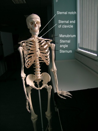
We looked at the bones of the neck, and ticked off the manubrium, the 1st rib and the clavicles as marking the inferior border of the neck, and the mandible and inferior parts of the skull as the superior border of the neck. I pointed out the mastoid process, which is part of the temporal bone in the skull. You saw the sternocleidomastoid muscle that was attached here in another anatomy station. We also briefly looked at the hyoid bone, which many of the strap muscles of the neck attach to.
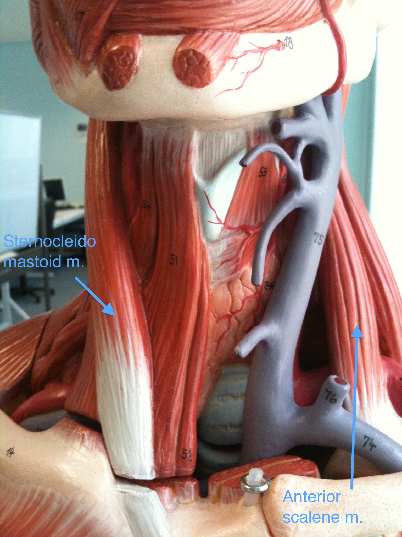
Digging deep looking at illustrations, Google’s body browser (you’ll need Google’s Chrome browser installed for this to work) and plastic models we found the anterior scalene muscle. It has siblings in the middle and posterior scalene muscles, and runs from the transverse processes of the cervical vertebrae (C3 – C6) down to the first rib. This muscle will elevate the ribcage, or flex the neck to either side.
The reason we picked out the anterior scalene muscle is because of other key structures that pass near to it. The brachial plexus and the subclavian artery passes between the anterior and middle scalene muscles, superior to the 1st rib on their way out to the arm. The phrenic nerve lies on the anterior surface of the anterior scalene muscle and then runs down into the thorax. The carotid sheath, containing the internal jugular vein, the common carotid artery and the vagus nerve, runs anterior and medial to the anterior scalene muscle too.
You now have a landmark to relate these important structures to, so you will be able to describe where they run as your knowledge of them develops. This will also help you find them.
We briefly touched on the blood supply to the trachea and upper oesophagus, linking it to the thyroid gland and the thyroid vessels (particularly the inferior thyroid arteries and the plexus of thyroid veins).
Finally we continued on from last week, where we looked at the arch of the aorta and the descending aorta in relation to the lungs. From the aortic arch 3 major blood vessels branch. The first branch is the brachiocephalic trunk. “Brachium” refers to the arm, and “cephalic” refers to the head, so this artery will supply blood to the arm and the head. It crosses across to the right side of the thorax behind the manubrium, and splits into the right subclavian and right common carotid arteries. The next branch from the arch of the aorta is the left common carotid artery (which is in the appropriate spot to be able to ascend directly up the neck because the aorta has already arched over to the left), and the third branch is the left subclavian artery.

Take a look at the torso models in the lab – some have the scalene muscles and blood vessels, some have the scalene muscles and nerves. Take a good look at how the arch of the aorta not only arches to the left, but also arches posteriorly to reach back to the posterior wall of the thorax. This is something that is often missed when just looking at in images in textbooks.
One of our models has a major flaw in the great vessels of the thorax and neck. if you didn’t find it on Monday have a good look at all of the torso models and see if you can find it.

Comments
5 responses to “Week 104: the neck”
Hi, I am a student nurse anesthetist and am constructing a presentation on difficult airways for an upcoming conference. Would you mind if I used the last picture on this page in my presentation?
Thank you for your consideration,
David
Hi David,
Yes, please do.
Sam.
Sam,
Thank you very much. It is a beautiful and very rich picture.
David
HI there
I have a client looking for a Flow Model for their students which is anatomically correct that shows the aortic arch and major arteries to the brain in order to demonstrate their products – do you know if such a thing exists and where we might find some? Please contact me on jo@thelittlebrandingcompany.co.uk
Sorry, no, I don’t know. If you want to accurately model the flow of blood through the vessels that’s quite a specific thing. Someone may have made one – maybe try searching scientific literature databases.