Adder by Wildlife Wanderer on Flickr.
Today we talked about the anatomy of the lymphatic vessels of the lower limb, and the mechanisms by which lymph is transferred from lower limb tissues back to the abdomen, thorax and circulatory system. We used examples of elephantiasis (and the filariasis worm), peripheral oedema, exercise recovery (after running or cycling racing) and snakebite.
As we’re midway through year 2 and students are close to having covered all of the human anatomy in the medical curriculum we were all aware of the purposes of the lymphatic system and some of the fine structure, but there was no harm in reviewing some of the smaller anatomical details to help understand the relations between structure and function here. For example, lymphatic vessels begin as small, open ended vessels into which fluid from a tissue can pass. This fluid is most likely to have come from the plasma of the capillary bed that perfuses the tissue, and not all of the fluid is collected on the venous side. Normally the fluid is returned to the systemic circulation by the lymphatic system but your foot is a long way away from your heart, so how does this work? The lymphatic system is a collection of vessels that drain into larger and larger vessels, but the flow is only in one direction, there is no pump attached directly to them, and the pressure within these vessels is very, very low.

Lymphatic capillaries (This file is licensed under the Creative Commons Attribution-Share Alike 3.0 Unported license).
We see examples of when the lymphatic system doesn’t work in peripheral oedema. Probably the most extreme version is infection of the lower limb by filariasis larvae though mosquito bite. These larvae develop into roundworms and become lodged in the lymph nodes of the lower limb. If the inguinal nodes become blocked the drainage of lymph is significantly impaired and fluid is retained in the lower limb. The legs swell to an enormous extent, hence the term, “elephantiasis”. If the scrotum is affected (by blockage of inguinal and iliac lymph nodes – remember that the lymphatic drainage of the testes themselves follows the gonadal vessels up into the abdomen and the para-aortic nodes) it can also swell as a hydrocele testis. Kill the larva and you prevent the problem from occurring. If it does occur the lymph nodes and the worms will probably have to be removed. This extreme example shows how important the lymphatic system is to normal, healthy body functions.

Wuchereria induced elephantiasis (This file is licensed under the Creative Commons Attribution-Share Alike 3.0 Unported license).
You’re more likely to see peripheral oedema in immobile or elderly patients. Pitting oedema is a swelling of the subcutaneous tissue that when pressed with your thumb remains indented for a few seconds. High blood pressure, histamine (makes capillaries leaky to proteins and immune cells), pregnancy (progesterone makes veins leaky), obesity, anaemia, deep vein thrombosis, hypothyroidism and some autoimmune disorders like lupus can cause pitting oedema. Why does it occur in immobile patients?
In muscles after athletic exertion ion imbalances cause fluid retention and tearing of sarcomeres, releasing fluid into the interstitial spaces (see also “Why do muscles hurt after exercise?). While we don’t see pitting oedema we do find swollen muscles, and we know that recovery seems to be aided by warming down and easy recovery movements like walking or cycling easily. Cold water or contrast water (hot – cold – hot – cold) therapy helps recovery, as do compression therapy (e.g. long compression socks and light massage. Why do these help?
Movement of the lower limb and contractions of the muscles cause the lymphatic system to drain fluid from the lower limb. The musculovenous pump aids venous drainage of the lower limb, and has the same effect on the lymphatics. Fluid that collects within tissues enters the first parts of the lymphatic vessels as the pressure in the tissue rises above about 1mmHg and small tethers linking the endothelial cells of the vessel wall are pulled, opening gaps and allowing fluid to pass in. As the pressure rises above 2mmHg within the vessel, either by muscular contraction around the vessel or by pulses of an adjacent artery the fluid is pushed along the lymphatic vessel, through valves that prevent back flow. Repeated contractions of lower limb muscles pushes fluid up the vessels, through lymph nodes and into the pelvis and abdomen.
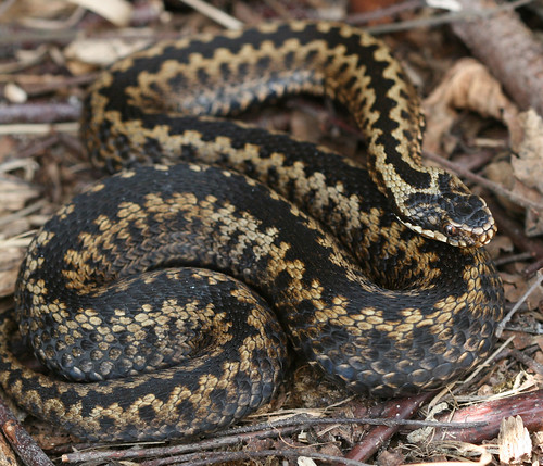
With oedema of the lower limb compression, activity and raising the limb all help drain the fluid from the limb by helping the lymphatic system. If you want to prevent flow of lymph up the lower limb, for example after a venomous snake bit to the foot (snake venoms tend to be large molecules that pass along the venous system) a compression bandage raising the pressure even higher will squash the lymphatic vessels, preventing flow. Be careful to keep the compression on the limb until in a medical treatment setting as removal will cause increased blood flow into the leg and increased flow of lymph out. Do the opposite of what you would do to treat oedema, and prevent movement, and don’t raise the limb.
There are superficial lymphatic vessels and deep lymphatic vessels in the lower limb, and as elsewhere they often follow veins. The superficial lymphatic vessels will follow the great saphenous vein back through the fossa ovalis to the femoral vein and the femoral triangle. The lymph will pass through superficial inguinal lymph nodes and then deep inguinal lymph nodes that are deep to the fascia lata. Flow continues through the external iliac nodes, the common iliac nodes and then the para-aortic nodes. Fluid from there can pass to the cistern chyli, thoracic duct and then return to the blood to the left subclavian vein near where the left internal jugular vein joins it.