 I wasn’t supposed to be teaching on Monday but one of our clinical colleagues was ill so it was a good job I could remember a little bit of kidney anatomy.
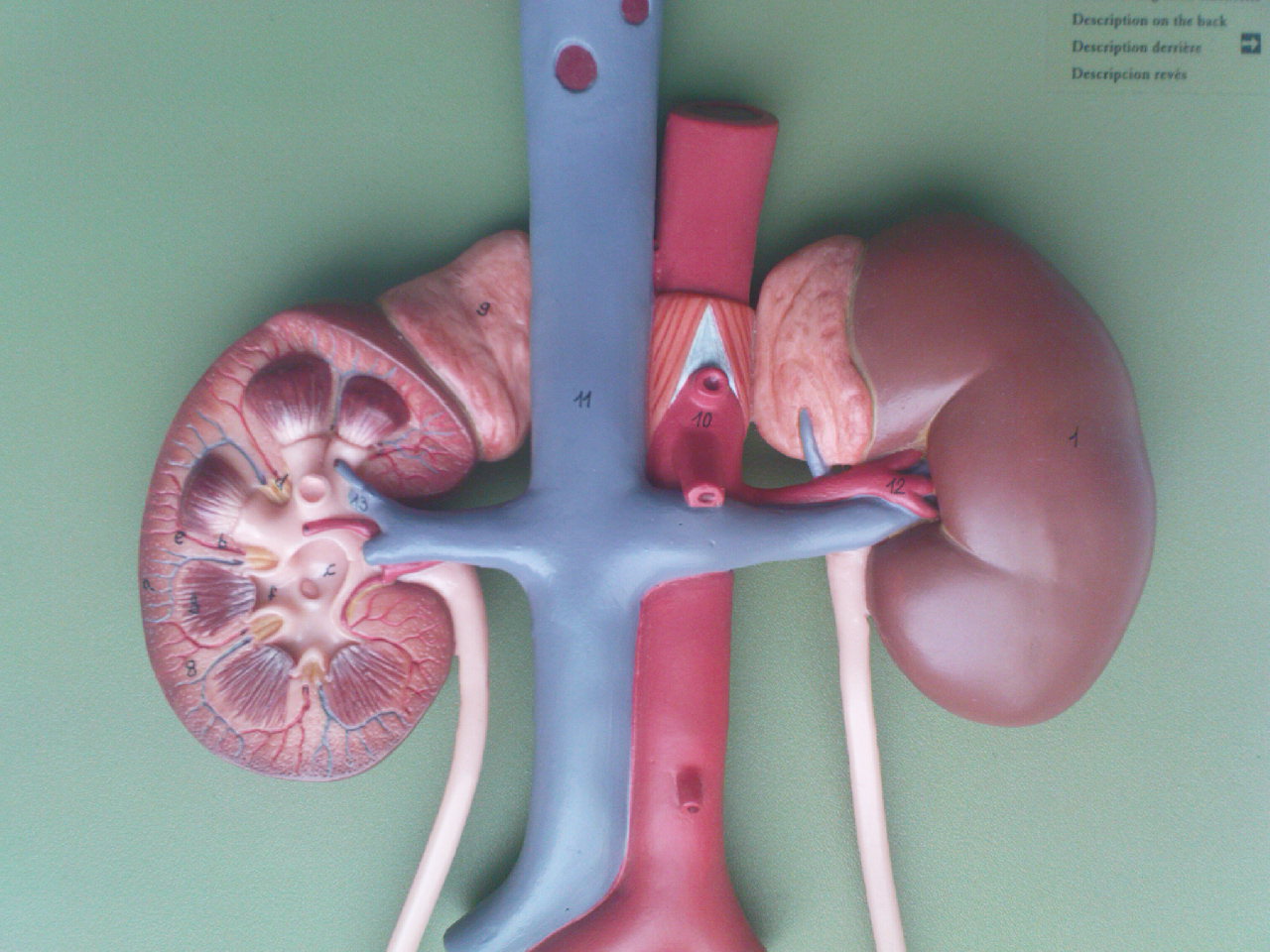
I briefly introduced the location of the kidneys, found retroperitoneally on the posterior abdominal wall. The right kidney is a little lower than the left, and they have different structures anterior to each (on the left and right sides of the abdomen, that is). I must remember to answer my own twitter question about this. We noted the aorta and the inferior vena cava and and the connections to & from the kidneys (renal arteries & veins).
Looking at the models and prosected material we noted that the left renal vein lies between the aorta and the superior mesenteric artery, and that this artery arises from the aorta just superior to this point. It is worth reviewing those three anterior branches from the abdominal aorta to link this renal anatomy with your GI tract learning. We also noted on one prosection that there were two right renal arteries and two left renal arteries in this particular case. Interesting!
We also took a look at the interior structure of the kidney, relating the cortex and the medulla to the histology and function of the kidney. In the cortex we would expect to see the glomerulus, the proximal convoluted and distal convoluted tubules, and in the medullary pyramids we should find the loops of Henle and the collecting ducts. The cortex is highly vascular, whereas the medulla is primarily composed of components of the urine collection system. Urine, we supposed, must collect at the apex of each medullary pyramid, and here we found spaces called minor calyces. Although we were looking at sections of the kidney we tried to imagine the roughly pyramidal shapes of the medullary segments, and that these pyramids were also arranged off the plane of section that we happened to be looking at (coronal). The models of the kidneys helped with this.
The minor calyces joined up and drained into larger spaces called major calyces, and these in turn drained into the renal pelvis from which the ureter began. Remember this separation of “blood” parts and “urine” parts of the kidney when you look at the embryological development of this organ.
Links:
Wikipedia has a labelled diagram of the internal parts of the kidney: link.
A study in 1986 by Gattone et al. attempted to trace the innervation of the kidney: link.
The National Kidney Foundation (USA) has lots of patient-friendly kidney info: link.