On Monday I spent most of the morning flexing my guns and poking my cubital fossa. Our aims were to look at the movements of the elbow joint, the muscles involved, and the important structures passing through this region, with particular regard to the cubital fossa. That video makes me feel a little bit ill. And I’m an anatomist.
We talked about the major movements of flexion and extension of the elbow joint, which you all were aware of, and mentioned pronation and supination of the forearm. I didn’t talk about the bones or the articulating surfaces in much detail as my clinical colleagues had pinched all the skeletons, and I’m sure they did a far better job of talking about the osteology than I would have done. Focusing on flexion and extension we identified the three main muscles involved: biceps brachii, brachialis and triceps brachii.
The biceps brachii muscle has 2 heads. The short head arises from the coracoid process of the scapula and the long head arises from the supraglenoid tubercle (lumpy bit above the glenoid cavity of the shoulder joint). The long head must pass through the shoulder joint, change direction and then run through the bicipital groove (or intertubercular sulcus) in the humerus. Have a brief look at the humerus and scapula in the anatomy lab to be clear on where these bony bits are.
The fibres of the biceps brachii muscle come together distally to insert into the radial tuberosity (another lumpy bit, on the radius). This means that not only is the biceps muscle a flexor of the elbow joint, but it can also powerfully supinate the forearm when the elbow joint is already flexed. Try this out next time you’re turning a screw or a bolt – feel biceps contract as you supinate the forearm and tighten the bolt. This also explains the different biceps flexing poses that bodybuilders may use to show off biceps (i.e. it’s bigger when the forearm is supine – get posing in front of the mirror to check this).
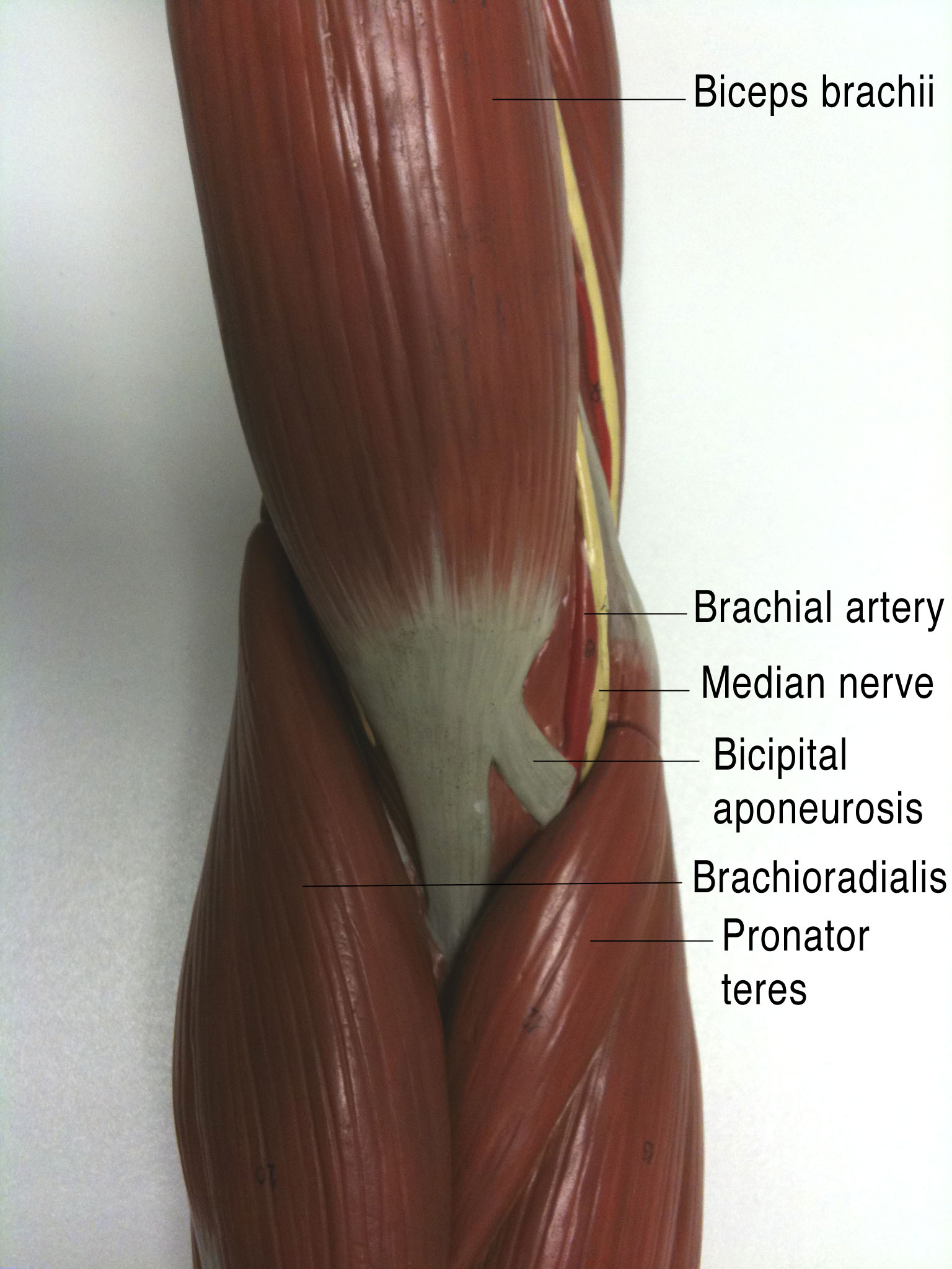
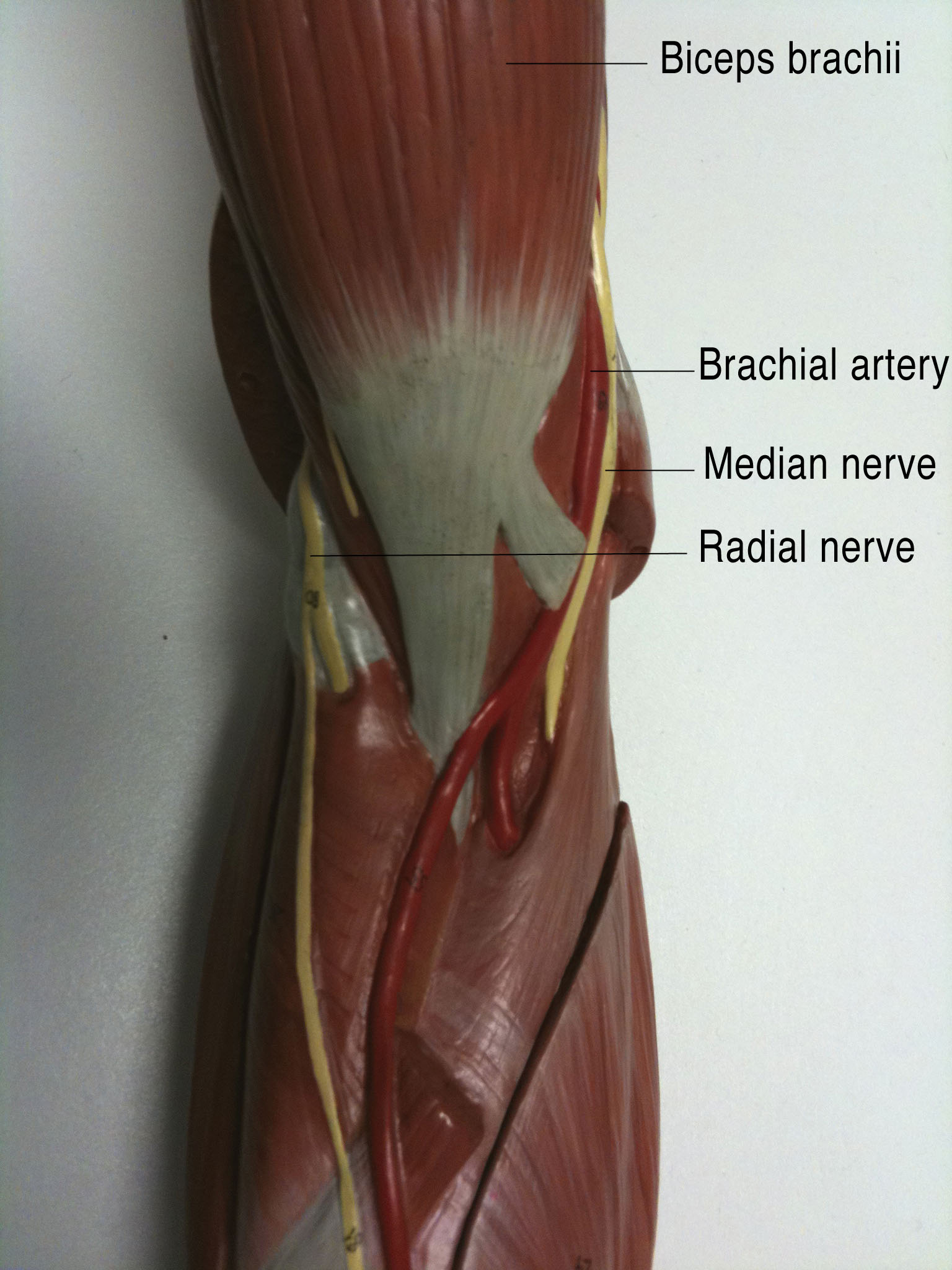
 The other thing we talked about with regards to biceps was that you can palpate not only the tendon inserting into the radius but also a weird, flat tendon medially. This is the bicipital aponeurosis (see me poking mine in the photo to the right), and connects the biceps to the deep fascia of the forearm. Clever, eh?
Don’t forget that as biceps brachii also crosses the shoulder joint it is also able to flex the glenohumeral (shoulder) joint, although it’s not great at this.
Deep to biceps we found the brachialis muscle arising from the humerus and passing across the elbow joint to insert into the tuberosity of the ulna. This muscle is flattened, and is the most powerful flexor of the elbow joint. The musculocutaneous nerve runs between brachialis and biceps brachii, innervating both muscles.
To extend the elbow joint we use one muscle: triceps brachii. By it’s name this must be a muscle of the brachium (upper arm) with 3 heads. The long head comes from the infraglenoid tubercle on the scapula (on the other side to the long head of biceps brachii), the medial head comes from the posterior surface of the humerus and the lateral head also comes from the posterior surface of the humerus, but a little more laterally. There’s a groove between the medial and lateral heads, and in this groove we find the radial nerve. This is the nerve of the posterior compartment of the arm and it is innervating the triceps muscle.
All of the muscle fibres of triceps come together to insert in the bony, sticky outy bit of the elbow (very sticky outy in my case, as some of you noticed): the olecranon. The olecranon is part of the ulna, so contraction of the triceps muscle pulls the olecranon and extends the elbow.
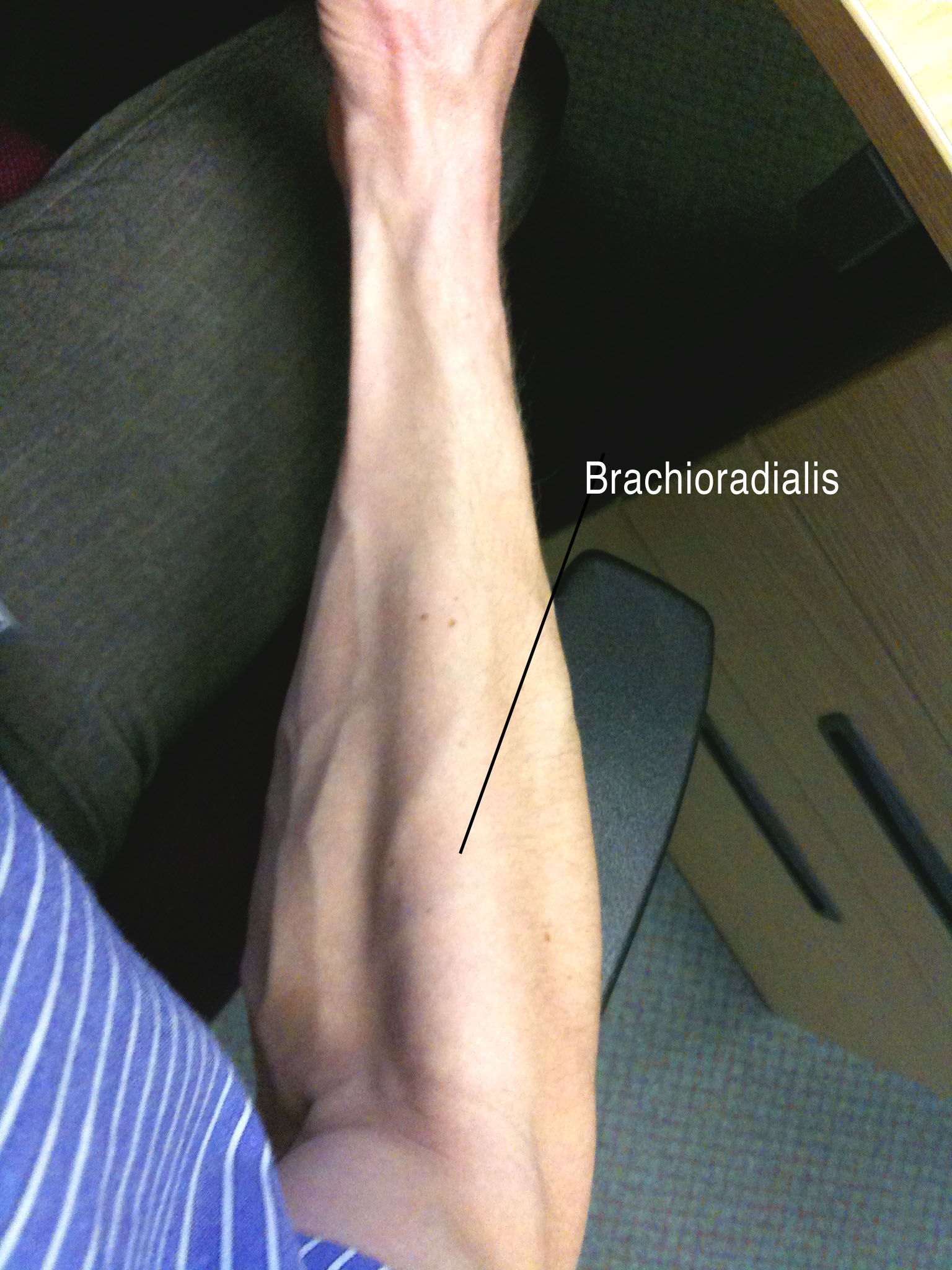 The next bit to look at was the cubital fossa. We had to find the brachioradialis and pronator teres muscles to find the lateral and medial borders, and add an imaginary line between the epicondyles of the humerus to add the proximal boundary. Brachioradialis is pretty easy to find on the lateral edge of the anterior supinated forearm, running from the humerus to the radius as the name implies. It is also innervated by the radial nerve, and when we lifted the brachioradialis muscle at the border of the cubital fossa we found the radial nerve twisting around the humerus to get to the anterior side.
Pronator teres was a little trickier to find, but it was clearly running diagonally across the proximal part of the forearm. Structures within the cubital fossa looked as though they would be protected by the biciptial aponeurosis.
What did we find in there? We saw the median nerve running (medially) alongside the brachial artery within the fossa itself (i.e. superficially to brachialis but deep to the fascia and skin) and noted that the median cubital vein, from which blood is commonly taken, was a superficial structure here. You remembered, of course, that this was where you listened to the brachial pulse when taking someones’s blood pressure.
After chatting about funny bones, we realised that we could now find all the major neurovascular structures of the upper limb at the elbow. The ulnar nerve is where we bang it (that made more sense during the discussion), the radial nerve is deep to brachioradialis, the median nerve is on the medial side of the cubital fossa with the brachial artery, and the musculocutaneous nerve peters out from between the biceps brachii and brachialis muscles as a cutaneous nerve, having done it’s “musculo” job.
Not bad for 25 minutes work.