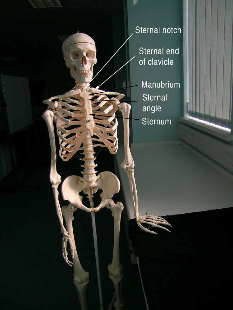
 When we looked at the surface anatomy of the thorax yesterday we focused on the bony bits anterior to the mediastinum. We found the sternal notch (or suprasternal notch, or jugular notch), the sternal ends of the clavicles, the manubrium, the sternal angle (or angle of Louis) and the sternum. Our aim was to look deep to these landmarks for major thoracic structures like large blood vessels and parts of organs and compile a table as we progressed.
So, what did we find?
Deep to the sternal notch almost all of the blood vessels have bifurcated to the left or right to avoid this weak spot. If we palpated the sternal notch we could press on the trachea, posterior to which on models we found the oesophagus and then the vertebral bodies themselves. We also found the inferior thyroid vein here, which is why you don’t use this region for an emergency tracheotomy, I guess.
Lateral to the sternal notch, the medial ends of the clavicles were found to overlie the brachiocephalic veins. In this region we saw the brachiocephalic vein on either side form as the large internal jugular vein from the neck joined with the subclavian vein. Deep to this we saw on the right side that the brachiocephalic trunk was deep to the right brachiocephalic vein here, but on the left side the left common carotid artery and left subclavian artery were independent of each other, having separately branched from the arch of the aorta (well, in all models except one!) On the right side we found that a little superior to the sternal ends of the clavicle the brachiocephalic trunk split to give the right common carotid artery and right subclavian artery. All of the large vessels in this region were well protected by the superficial muscles of the neck and the bones of the clavicle and manubrium. Rarely, the vagus nerves and phrenic nerves were discovered descending through this region on either side (the vagus nerves were passing with the carotid arteries, and the phrenic nerves were upon the anterior scalene muscles (look at the images in those links) – you might need to hunt again for these).
 Moving inferiorly, on some models we saw how the superior vena cava formed from left and right brachiocephalic veins deep to where the 1st rib joined with the manubrium. Posterior to the manubrium was the arch of the aorta.
At the sternal angle (where the manubrium meets the sternum, very palpable) we could form a plane across to the intervertebral disk between vertebrae T4 and T5 (the transverse thoracic plane). Superior to this plane was the superior mediastium, and inferior to this plane was the inferior mediastinum (which was further split into anterior, middle and posterior mediastinum by the heart and pericardium). The plane cut through the start of the aortic arch and its end, where it becomes the thoracic aorta. We also found the carina, where the trachea bifurcates into the main bronchi, and found that this is a great place to start counting ribs from (rib 2 is here).
The right side of the heart was found deep to the sternum, and we noted that if you drew a line from the middle of the left clavicle down the thorax, perpendicular to the floor, the apex of the heart’s left ventricle extended this far. This line is a midclavicular line.
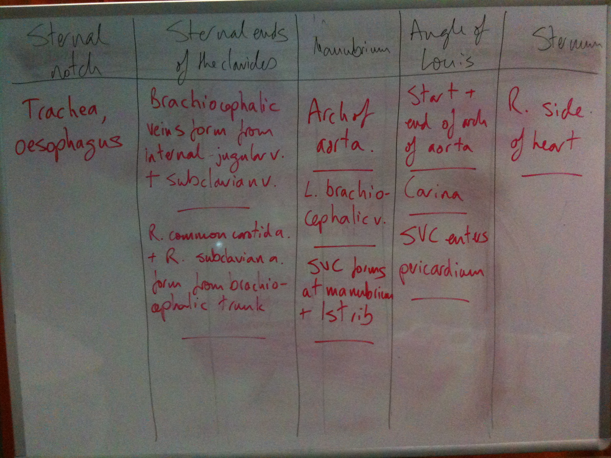
Here’s one of the tables that we compiled: