 In our first anatomy session on Monday we began the year by introducing the abdomen. It’s a good place to start as most of the anatomy here is fairly straightforward and it gets you thinking in three-dimensions. The concept of the peritoneum is the toughest part to understand, as are the mesentery, mesocolon, omenta and sacs that are formed by it’s folds and spans between organs.
In my session I spoke a little about anatomy teaching and learning, and how to best use the small group format. Hopefully we’ll be able to look at models, prosections and whatnot together as a small group, and we’ll change the format a little from week to week (most weeks I’ll talk to you, but some weeks I’ll get you to find and show each other the structures that we’re interested in, for example). There’s a huge amount of human anatomy to learn, and I guess the best ways for most people to store & recall all this information are to engage with and use the stuff that you’re learning. Use it or lose it, as they say.
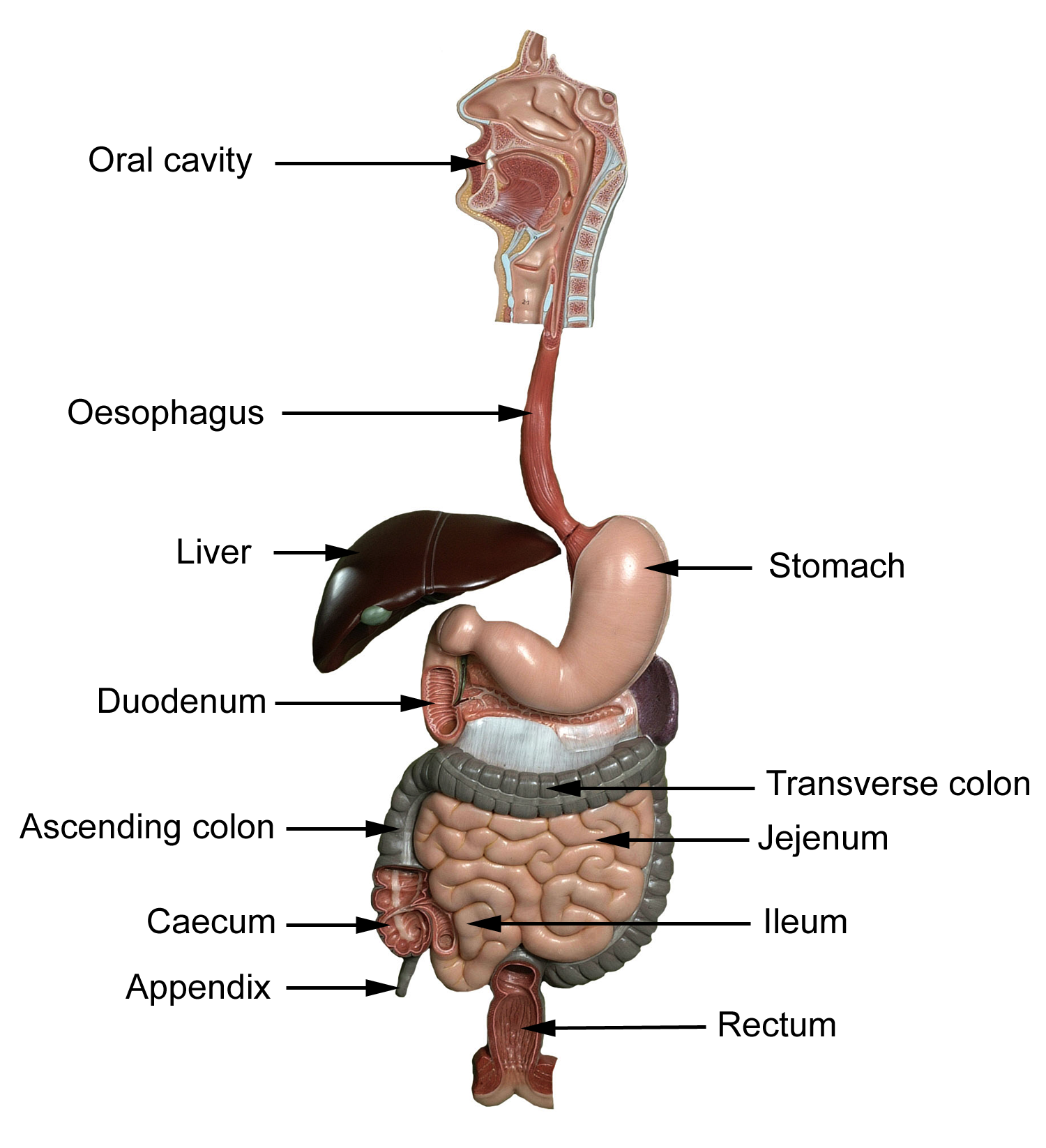
So, I was given (and passed on to you) the learning objective of, “identify the different parts of the small and large intestines, and describe their locations in the abdomen and list the anatomical differences between them.”
We started at the top, and worked down quickly to the first part of the small intestine. We found the start of the duodenum as it left the pylorus of the stomach and noted that the duodenum is fairly well fixed in place (certainly when compared to the rest of the small intestine) and is partly retroperitoneal and held against the posterior abdominal wall. We saw the C-shaped curve that the duodenum makes, and saw that it had 4 parts: superior, descending, inferior and ascending. We also saw the reason for the duodenum’s curve – it has the head of the pancreas nestled into it, and the pancreas ducts its exocrine secretions into the duodenum here, into the descending (or 2nd) part, aiding digestion.
So one of the functions of the small intestine is continued digestion then.
Looking inside the duodenum we saw that the internal surface was very corrugated, and we could see that the rest of the small intestine was very long (about 6m) and very folded. In Paul Griffiths’ histology bit you saw that the histology of the small intestine showed even more surface area optimisation. All this surface area increases the ability for the small intestine to pass molecules across cells and into the blood stream. So another function of the small intestine is absorption of nutrients (but you probably already knew that, right?)
The duodenum starts about the level of the first lumbar vertebra (L1), drops down to the level of L3, and then moves across to the left side and rises a little to the level of L2. From here the jejunum begins, and coils and folds, held in place by its mesentery. Jejunum becomes ileum (spelling is important), and the ileum ends the small intestine in the lower right quadrant where it meets the large intestine. The jejunum probably makes up about 40% of the small intestine, and the ileum the other 60%. Some differences between the jejunum and ileum are noted here.
The remaining contents of the small intestine enter the large intestine at the caecum, a short pouch that starts off the large bowel from which the appendix hangs. The ileocaecal valve prevents contents from passing back into the ileum when the peristaltic contractions of the colon push its contents up through the ascending colon.
The ascending colon is usually retroperitoneal and well fixed in place, and the large intestine makes a left turn at the right colic flexure (or hepatic flexure) where it nestles into the liver. The transverse colon is much more free to move as it has a mesocolon (like the small intestine’s mesentery) attaching it to the posterior abdominal wall. At the left side the colon makes another turn, and this turn is called the left colic flexure (or splenic flexure). It descends as the descending colon (retroperitoneal), becomes the sigmoid colon (which has a mesocolon), and continues inferiorly but also posteriorly, moving towards the pelvis. The sigmoid colon becomes the rectum, which stores faces for removal through the anal canal.
The large intestine has a larger diameter than the small intestine (some naming in anatomy really is sensible) and one of its functions is to remove water from its contents and compact the leftovers to form faeces (more functions here).
That takes us to the bottom. Sorry, to the end. Take a look at the large bowel (large intestine, colon) and find the taenia coli (smooth muscle strips), the haustrae (pouches), and the omental appendices (fat-filled pouches of peritoneum) too. They’ll tell you that you’ve found large, and not small, intestine.