 Hey! I hadn’t taught for a while so it was good to start again, in hopefully a helpfully interactive way as we all looked at sliced and whole kidneys.
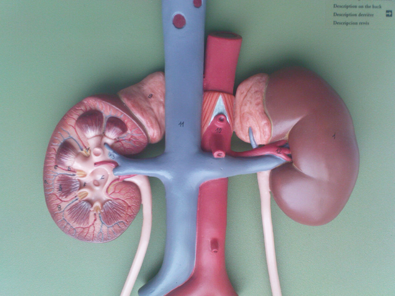
In the session we talked about the cortex and the medulla, pointing out that while the cortex is outermost it also surrounds medullary (or renal) pyramids so there are renal columns (cortex) between pyramids through which blood vessels pass. We tried to relate the functional histology of the kidney that you saw in Paul Griffiths’ session to the gross anatomy that we had in the lab. We talked about the glomerulus, the proximal convoluted and distal convoluted tubules in the cortex and mentioned the loops of Henle and the collecting ducts in the medullary pyramids. Urine, we supposed, must collect at the apex of each medullary pyramid, and here we found spaces called minor calyces.
The minor calyces join up and drain into larger spaces called major calyces, and these in turn drain into the renal pelvis from which the ureter begins. Remember this separation of “blood” parts and “urine” parts of the kidney when you look at the embryological development of this organ.
We talked about autonomic innervation and the routes that sympathetic nerves might take to get to the kidneys (sympathetic trunk, splanchnic nerves, prevertebral ganglia). While sympathetic innervation will cause vasoconstriction here, so reducing urine output we reminded ourselves that most of kidney function is controlled hormonally.
The blood supply to the kidneys is clear, but we paid attention to the level at which the renal arteries pass to the kidneys (remember the superior mesenteric artery here) and looked at the relationship between the renal arteries and veins (the left renal vein lies anterior to the aorta). We made sure that we could differentiate between a ureter and a blood vessel at the hilum of a kidney specimen.
More information:
Wikipedia has a labelled diagram of the internal parts of the kidney: link.
A study in 1986 by Gattone et al. attempted to trace the innervation of the kidney: link.
The National Kidney Foundation (USA) has lots of patient-friendly kidney info: link.